Renal Physiology
Danielle E. Soranno, M.D.
Assistant Professor
University of Colorado
Children's Hospital Colorado
13123 East 16th Ave
Aurora CO 80045
→ Enlace a la versión en español
Danielle E. Soranno, M.D.
Assistant Professor
University of Colorado
Children's Hospital Colorado
13123 East 16th Ave
Aurora CO 80045
→ Enlace a la versión en español
Overview of renal function:
The kidneys play a critical role in the normal health and development of children. The nephron is the functioning unit of the kidney, with each kidney containing ~500,000-1,000,000 nephrons. Cumulatively, the basic processes of the nephron are 1) filtration, 2) reabsorption, and 3) secretion.
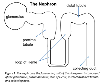
Each nephron has 5 main parts: 1) the glomerulus, 2) the proximal tubule, 3) the loop of Henle, 4) the distal convoluted tubule, and 5) the collecting ducts (Figure 1). The glomeruli (‘filters’) have both charge and size selectivity and filter plasma across the endothelial (capillary), glomerular basement membrane, and epithelial (podocyte) layers. (See below regarding assessment of filtration.) The proximal tubule (‘workhorse’) is where approximately 85% of reabsorption occurs – including sodium, water, amino acids, and glucose. The loop of Henle allows for dilution and subsequent concentration of urine via the counter current multiplier. The distal convoluted tubule contains the macula densa which is critical for juxtaglomerular feedback with subsequent autoregulation of renal blood flow. Autoregulation allows for a fairly constant glomerular filtration rate (GFR) under normal physiological circumstances. Finally, the collecting ducts respond to anti-diuretic hormone (ADH), which allows for concentration of urine, as well as aldosterone which allows for secretion of potassium.
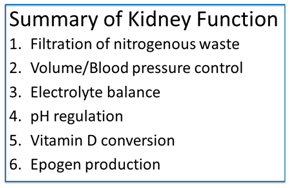
Beyond filtration, the kidneys also control volume and blood pressure (via both response to anti-diuretic hormone as well as the renin angiotensin aldosterone system), regulate electrolyte balance, help regulate serum pH, convert inactive 25-hydroxy vitamin D to active 1-25-dihydroxyvitaminD, and secrete epogen.
Assessing renal function:
Despite the complexity of filtration, reabsorption and secretion along the nephron, assessment of nephron function can be simplified into 1) filtration (glomeruli) and 2) tubular function.
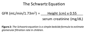
Glomerular filtration rate (GFR) is the rate at which plasma is cumulatively filtered through the nephrons. After filtration, most of the filtered load is reabsorbed along the nephron. In pediatrics, the Schwartz equation is most commonly used to estimate GFR in children[1]. The Schwartz equation (Figure 2) is based on serum creatinine as well as the child’s height. While a normal GFR in adults is ~120 mL/min/1.73m2, in pediatrics a normal GFR varies depending on a child’s age. Typically, a full-term neonate’s initial GFR is only ~30 mL/min/1.73m2 while a premature neonate’s initial GFR can be even lower ~15 mL/min/1.73m2. Regardless of gestational age, GFR roughly doubles within 2 weeks of age because of decreasing vascular resistance with a resultant increase in renal blood flow and perfusion. GFR then continues to increase (corrected for body surface area) and reaches adult norms about about 19 months of age.
Tubular function can be assessed via electrolyte balance as well as the ability to concentrate and dilute urine. Tubulopathies can often be successfully diagnosed via interpretation of electrolyte abnormalities[2]. For example, tubular wasting of sodium, phosphorus, potassium, amino acids and glucose should precipitate an evaluation for Fanconi syndrome. Fanconi syndrome occurs when there is a global defect in proximal tubular function with resultant urinary losses of the filtered load that would normally be reabsorbed[3]. Children with Fanconi syndrome typically present with failure to thrive.
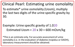
As stated previously, urinary concentration occurs in the collecting ducts in the presence of, and renal response to anti-diuretic hormone (ADH). The concentration gradient that allows for concentrating the urine is set up in the loop of Henle, which first dilutes urine. Urine can be diluted to ~50 mOsm/kg and concentrated to ~1200 mOsm/kg. Full concentrating ability takes several months to develop, and a typical full-term neonate can only concentrate their urine to ~500 mOsm/kg. This concentrating defect of infancy is a result of an immature counter current multiplier as well as relative tissue insensitivity to ADH[4].
In general, if 1) serum electrolytes (sodium, potassium, phosphorus, calcium, bicarbonate) are within normal limits, 2) the urinalysis indicates that the child can concentrate or dilute their urine, and 3) there is no tubular wasting of glucose or amino acids/protein, then tubular function can be assessed as normal.
Acute Kidney Injury
Acute kidney injury (AKI) refers to a sudden decrease in glomerular filtration rate. AKI is typically reversible, however there is a growing appreciation that AKI can lead to chronic kidney disease (CKD). There are several staging classifications of AKI[5] such as the p-RIFLE criteria, which stratifies AKI in children between “Risk, Injury, Failure, Loss and End-Stage.” The RIFLE criteria are based upon urine output and rise in serum creatinine[6].
In the setting of AKI, it is important to understand that a rise in creatinine typically appears only after significant renal dysfunction has begun, and often several days following the initial insult - well into the process of kidney injury.
A typical starting point to determine the etiology of AKI includes an assessment of prerenal, intrinsic renal, or obstructive processes. Obstruction can be ruled out via imaging, typically via renal and bladder ultrasound. Prerenal and intrinsic etiologies of kidney injury will be discussed herein.
Prerenal azotemia refers to a decrease in GFR secondary to poor renal perfusion, often in the setting dehydration or volume loss. Patients who are prerenal will often exhibit oliguria (urine output <0.5 mL/kg/hr in children, <500 mL/day in adults) and will demonstrate signs and symptoms of dehydration (tachycardia, dry mucus membranes, hypotension, increased skin turgor). Assessment of a urinary fractional excretion of sodium (FENa) will be very low (<1%) (Table 1), indicating that the renal tubules are appropriately reabsorbing sodium. Blood urea nitrogen (BUN) is usually elevated in prerenal azotemia. This is a result of an increase in reabsorption of sodium in the proximal tubule which results in a passive reabsorption of both water and urea nitrogen. Thus, the patient’s serum BUN to creatinine ratio will be elevated in prerenal azotemia. The treatment of prerenal azotemia is intravascular volume repletion, typically via normal saline boluses. After attaining euvolemia, it is important not to continue to bolus with excessive fluids, as prerenal azotemia can lead to acute tubular necrosis (see below) if the injury is long-standing, and in such cases, the patient may not be able to make urine. In this case, excessive fluid resuscitation can lead to hypervolemia with resultant hypertension, edema, and respiratory distress.
Intrinsic renal failure refers to AKI that is not secondary to prerenal or obstructive causes. It is most helpful to refer to the construct of the nephron when considering intrinsic AKI as this correlates with the various etiologies: 1) vascular causes; 2) glomerulonephritis; 3) acute tubular necrosis; and 4) acute interstitial nephritis.
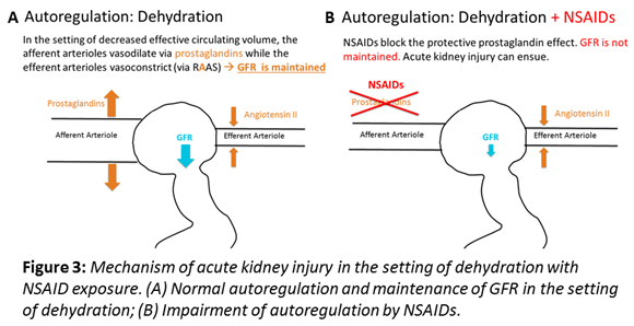
1. Vascular causes of AKI are quite common and include any pertubation in blood flow to the kidney – most commonly secondary to medications such as NSAIDs or cyclosporine/tacrolimus, which cause vasconstriction of the afferent arterioles, or ACE-inhibitors/ARBs such as lisinopril, which cause vasodilation of the efferent arterioles[7]. For this reason, it is critical to remember that NSAIDs, including ketorolac and ibuprofen are completely contraindicated in any patient who is dehydrated, see Figure 3[8].
Thrombotic microangiopathies (TMA) such as the hemolytic uremic syndrome can also be classified as a vascular cause of AKI. In TMA, platelet rich thrombi form in the capillary loops of the glomeruli, leading to a decrease in GFR.
of the urine sediment showing red blood cell casts. The hallmarks of acute glomerulonephritis include hypertension, edema and AKI. The differential diagnosis of glomerulonephritis can be divided into two main categories based on serum complement levels – hypocomplementemic or normocomplementemic. Hypocomplementic GNs include post-infectious GN, systemic lupus erythematosis, and membranoproliferative GN. Normocomplementemic GNs include IgA nephropathy, Alport’s syndrome, and the ANCA vasculitides.
Treatment of acute GN depends on the underlying cause, and other than in the self-resolving 2. Glomerulonephritis (GN’s) can typically be diagnosed quite readily with evaluation illness of post-infectious GN, most GNs require treatment with immunosuppressive therapy.
3. Acute tubular necrosis (ATN) is the most common cause of AKI. ATN is usually caused either by ischemia-reperfusion injury, or exposure to nephrotoxic medications. Patients with ATN are typically oliguric and their urine sediment may demonstrate muddy brown or granular casts. In contrast to prerenal azotemia, patients with ATN have a FENa >1% (Table 1).
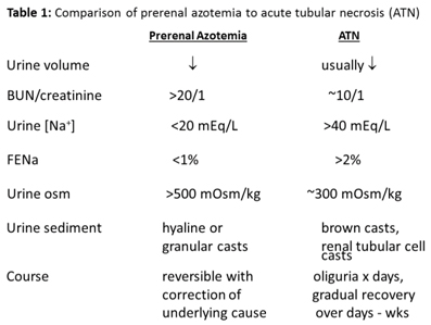
have a FENa >1% (Table 1). Treatment of ATN includes renal rest – maintaining euvolemia and avoidance of nephrotoxic medications. In oliguric patients, strict attention to fluid balance is necessary to maintain euvolemia. Recovery from ATN may take days to weeks, and often the oliguria transitions to polyuria with a persistently elevated serum creatinine. When the patient becomes polyuric, it is critical to maintain adequate hydration. Until the renal tubules recover, the patient is unlikely to be able to concentrate their urine, and as such they are at risk for developing prerenal azotemia with a potential secondary renal hit if intake is not increased to keep up with output.
A good rule of thumb when dealing with such patients is to obtain daily weights, strict in’s and out’s and to maintain the euvolemic patient on 1/3 x their maintenance fluid plus 1:1 replacement of urine output. The 1/3 x maintenance approximates the patient’s insensible fluid losses, while the replacement of urine output will work to maintain euvolemia in both oliguric and polyuric patients. In patients who are npo, typical fluids can be 5% dextrose 0.45% normal saline, however serum electrolytes should be followed closely. Polyuric patients may need addition of potassium to their intravenous fluids. Patients who have loose stools may need additional repletion for stool losses, including the addition of bicarbonate to the fluids.
4. Acute interstitial nephritis (AIN) is an underappreciated cause of AKI. AIN is often triggered by a reaction to an offending medication and is sometimes referred to as allergic interstitial nephritis. AIN may also be triggered by a viral infection, however most causes of AIN are idiopathic. While historically, urine was often sent for evaluation of eosinophils, eosinophiluria is neither sensitive nor specific for diagnosing AIN. As such, AIN can be diagnosed clinically in the setting of a patient with non-oliguric AKI with a bland urine sediment. Treatment of AIN includes cessation of the offending medication when clinically possible. AIN can also be treated with steroids, however, if the cause is medication-related, kidney injury may return once the steroids are discontinued. A rare but associated disease is tubulointerstitial nephritis and uveitis (TINU)[9]. These patients have both AIN and uveitis. TINU is often responsive to steroids, however, the uveitis requires ophthalmogic evaluation and treatment separate and in addition to renal evaluation and treatment.
Clinical pearl: Patients with an intra-abdominal urinary leak can inappropriately be diagnosed with AKI. In these patients, their serum creatinine rises secondary to reabsorption of urine creatinine across the peritoneal membrane with a resultant rise in serum creatinine. In this case, serum creatinine no longer reflects renal filtration. The diagnosis should be suspected in any patient with new development of ascites in the setting of a rising creatinine, who is at risk for urinary leak (i.e. recent surgical procedure).
Indications for dialysis:
There is no current treatment for AKI other than supportive care. Renal replacement therapy may be required if optimal medical management does not suffice. Indications for initiating acute dialysis include volume overload, electrolyte derangements, metabolic acidosis or uremia.
Chronic Kidney Disease
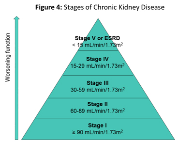
Chronic kidney disease (CKD) refers to either laboratory or radiographic evidence of kidney disease that progresses beyond 3 months. There are five stages of CKD, based on estimated glomerular filtration rate (Figure 4). In children with CKD, a modified version of the Schwartz equation, known as the “Bedside CKiD Equation” is used to estimate their GFR[1, 10]. Unlike adult patients who are often followed by their primary care provider throughout the early stages of CKD, all children with CKD should be followed by a pediatric nephrologist for subspecialty care[11]. Particular attention is paid to the growth and development of children with CKD. The frequency of nephrology follow-up correlates with the stage of CKD. Of note, GFR reaches its max at around 2 years of age, making it impossible to stage CKD in children less than 2 years of age accurately.
Management of Chronic Kidney Disease
Unfortunately, there is no cure for CKD. However, early referral to a pediatric nephrologist can help with management of co-morbidities and delay progression of disease. Routine CKD management includes assessment of staging, blood pressure control, as well as monitoring for anemia, growth delay, and metabolic bone disease.
Blood pressure control: Tight blood control to the 50th% for age, sex and percentile height has been shown to delay progression of CKD[12]. ACE-inhibitors and ARBs are often the preferred agent to control blood pressure as they also protect against glomerular hyperfiltration. For this reason, however, they are contraindicated in patients with borderline stage IV and V GFR or in patients who tend to have hyperkalemia.
Anemia of chronic kidney disease: Children with CKD have several reasons to be anemic – including epogen deficiency and increased hepcidin levels which leads to poor utilization of iron stores[13]. Patients with CKD should have routine CBC’s to monitor for the development of anemia. If and when they become anemic, iron stores should be checked and normalized. Patients who remain anemic despite adequate iron stores should be started on erythropoiesis stimulating agents such as epoetin alfa or darbopoetin alfa[11].
Metabolic bone disease (MBD): Formerly referred to as secondary hyperparathyroidism, metabolic bone disease encompasses not only secondary hyperparathyroidism, but also bone mineralization and vascular calcifications. MBD is often the most challenging aspect in managing children with CKD, and is unfortunately the reason for a significant amount of morbidity and mortality in our pediatric patients with CKD. MBD leads to increased cardiovascular disease, the leading cause of death in patients with CKD. Indeed, a pediatric patient with end stage renal disease has the cardiovascular risks of a patient in their 70’s[14]. Management includes maintaining adequate stores of 25-hydroxy vitamin D, maintaining normocalcemia and normal serum phosphorus levels, and supplementation of active 1-25-dihydroxyvitamin D[15, 16]. It is often quite difficult for patients to comply with the prescribed phosphorus binders which are taken with meals, and often cause gagging and discomfort. This is particularly difficult in young children, with whom mealtime can already be quite challenging.
Growth: Children with CKD often have difficulty with growth and development. A multidisciplinary team including a renal dietician is critical for proper monitoring of nutritional intake and growth. Unlike adults with CKD, children do not have a reduced protein limit, as they require protein for growth. We therefore recommend children with CKD obtain the recommended daily allowance of protein. Linear growth is particularly challenging because of a relative tissue insensitivity to growth hormone. Infants and toddlers with CKD often require gastrostomy tubes for either medication administration and/or supplemental formula feeds. Special renal formulas that support growth and nutrition while limiting potassium and phosphorus are often required. Supplemental growth hormone is indicated in patients not growing well linearly despite adequate caloric intake. Unfortunately, many families decline growth hormone treatment in order to save their child a daily injection, however quality of life studies have demonstrated that adult height positively correlates with quality of life[17, 18].
Renal replacement therapy (RRT)
Children who are progressing to stage V CKD require evaluation for renal replacement therapy. RRT can be in the form of either peritoneal dialysis (PD), hemodialysis (HD), or renal transplantation. For patients who have a potential living donor, pre-emptive transplantation may be a suitable and often desirable option. Evaluation of a living donor make take several months, so such a preemptive transplantation should begin while the patient is still in stage IV CKD. Similarly, introducing the concepts of dialysis and transplantation at an earlier stage, such as stage III often benefits the family and patient so they can begin to think about dialysis modalities versus preemptive transplantation.
The term ‘dialysis’ refers to two separate mechanisms: 1) diffusion of solutes across a semipermeable membrane, and 2) removal of fluid via ultrafiltration.
Peritoneal Dialysis (PD):
In peritoneal dialysis, dialysate is placed in the peritoneal cavity. The peritoneal membrane acts as the semipermeable membrane and allows solutes to flow across and down their concentration gradient. Ultrafiltration is controlled via the addition of dextrose to the dialysate fluid. By increasing the dextrose concentration to the dialysate, the osmotic gradient increases, thereby pulling more fluid from the patient into the peritoneal cavity. The fluid is then drained from the peritoneum. Most pediatric patients undergo home PD at night while they are asleep. In this way, they undergo several cycles, typically anywhere from 6-12 cycles. The PD prescription therefore includes the fill volume, dwell time, and dialysate to be used (including the dextrose concentration). The additional volume that is drained during each cycle drain is tallied and the total accumulated over the treatment is referred to as the ultrafiltrate (UF) volume. Of note, patients may retain fluid during PD and this typically indicates they might have been hypovolemic at the initiation of the treatment. Home PD requires extensive training of the caregivers with frequent communication with their nephrology center. Caregivers are expected to keep daily logs of pre- and post- treatment weights, blood pressures, dialysate used and UF obtained. Many patients have a sliding scale prescription such that the dextrose concentration can be adjusted based on their pre-dialysis weight and blood pressure.
Peritonitis is the main risk in patients who undergo PD. Caregivers are taught to recognize the signs and symptoms of peritonitis and are also trained to obtain a peritoneal effluent for cell count and culture prior to initiating intraperitoneal antibiotics. PD patients on antibiotics should always receive anti-fungal prophylaxis to prevent fungal peritonitis. Fungal peritonitis is both difficult to diagnosis as well as treat, and unfortunately requires removal of the PD catheter and has a high risk of scarring the peritoneal membrane.
Membrane failure can result from fungal peritonitis, recurrent bacterial peritonitis, or may be a result of glycosylation of the membrane from prolonged exposure to the dextrose in the dialysate. Routine peritoneal membrane tests are performed by their nephrologist to assess membrane characteristics and to help adjust PD orders over time.
Hemodialysis (HD):
In hemodialysis, the patient’s blood is circulated thru a dialysis machine containing an HD filter. Most filters are now hollow-fiber capillary membranes. Each fiber is hollow and allows blood to flow thru the tube, in countercurrent to dialysate flowing around the fibers. This design allows for a large surface area where-in the fibers are the semi-permeable membrane that allow for flow of solutes across the membrane and down their concentration gradient. UF is attained via negative pressure applied by the dialysis machine. The UF rate is limited by patient size, blood pressure and symptoms. Dialysis access can be challenging in pediatric patients. Often, fistula access is limited by patient size and vasculature. As a result, many pediatric patients have the additional risk of central line access, known to have an increased risk of infection above fistulas and grafts[11].
The choice of dialysis modality is made based on a combination of the child’s current medical status, underlying disease, family support and often, geographic location (i.e. distance from a hemodialysis center that will treat children). Past episodes of peritonitis or surgical procedures that have left adhesions may limit the option to pursue peritoneal dialysis. Certainly, peritoneal dialysis requires more social stability as well as time, energy and commitment from the child’s caregivers. Despite these additional responsibilities, PD is often a preferred choice as it allows the child to continue to go to school. Alternatively, hemodialysis (HD) is a good alternative for patients who have either failed PD, or who’s family may be unable to provide PD at home.
Regardless of the dialysis modality chosen, the goal of every pediatric nephrology program is to successfully tranplant their pediatric patients with ESRD. The life expectancy of children with ESRD on dialysis is 50 years shorter than healthy age-matched cohorts, compared to 15 years shorter following transplantation[14]. Transplantation evaluation includes psychosocial evaluation and at times, transplantation is delayed or put on hold if the child is not interested or is deemed to be a poor candidate based on non-compliance with prescribed therapies.
1. Schwartz, G.J. and D.F. Work, Measurement and estimation of GFR in children and adolescents. Clin J Am Soc Nephrol, 2009. 4(11): p. 1832-43.
2. Ariceta, G. and J. Rodriguez-Soriano, Inherited renal tubulopathies associated with metabolic alkalosis: effects on blood pressure. Semin Nephrol, 2006. 26(6): p. 422-33.
3. Fanconi, A. and A. Prader, [Primary tubulopathies. I. A case of idiopathic renal tubular acidosis (Albright's syndrome)]. Helv Paediatr Acta, 1961. 16: p. 609-21.
4. Cadnapaphornchai, M.A., et al., Neonatal Nephrology. Merenstein & Gardner's Handbook of Neonatal Intensive Care, ed. S.L. Gardner, et al. 2015.
5. Sutherland, S.M., et al., AKI in hospitalized children: comparing the pRIFLE, AKIN, and KDIGO definitions. Clin J Am Soc Nephrol, 2015. 10(4): p. 554-61.
6. Akcan-Arikan, A., et al., Modified RIFLE criteria in critically ill children with acute kidney injury. Kidney Int, 2007. 71(10): p. 1028-35.
7. Goldstein, S.L., et al., Electronic health record identification of nephrotoxin exposure and associated acute kidney injury. Pediatrics, 2013. 132(3): p. e756-67.
8. Musu, M., et al., Acute nephrotoxicity of NSAID from the foetus to the adult. Eur Rev Med Pharmacol Sci, 2011. 15(12): p. 1461-72.
9. Yitzhaki, P., [Tubulo-interstitial nephritis and uveitis syndrome--TINU syndrome]. Harefuah, 2000. 138(5): p. 360-2, 423.
10. Wong, C.J., et al., CKiD (CKD in children) prospective cohort study: a review of current findings. Am J Kidney Dis, 2012. 60(6): p. 1002-11.
11. Lamb, E.J., A.S. Levey, and P.E. Stevens, The Kidney Disease Improving Global Outcomes (KDIGO) guideline update for chronic kidney disease: evolution not revolution. Clin Chem, 2013. 59(3): p. 462-5.
12. Group, E.T., et al., Strict blood-pressure control and progression of renal failure in children. N Engl J Med, 2009. 361(17): p. 1639-50.
13. Atkinson, M.A., et al., Hepcidin and risk of anemia in CKD: a cross-sectional and longitudinal analysis in the CKiD cohort. Pediatr Nephrol, 2015. 30(4): p. 635-43.
14. Mitsnefes, M.M., Cardiovascular disease in children with chronic kidney disease. J Am Soc Nephrol, 2012. 23(4): p. 578-85.
15. Wesseling-Perry, K. and I.B. Salusky, Chronic kidney disease: mineral and bone disorder in children. Semin Nephrol, 2013. 33(2): p. 169-79.
16. Kumar, J., et al., Prevalence and correlates of 25-hydroxyvitamin D deficiency in the Chronic Kidney Disease in Children (CKiD) cohort. Pediatr Nephrol, 2016. 31(1): p. 121-9.
17. Goldstein, S.L., A.C. Gerson, and S. Furth, Health-related quality of life for children with chronic kidney disease. Adv Chronic Kidney Dis, 2007. 14(4): p. 364-9.
18. Goldstein, S.L., et al., Quality of life for children with chronic kidney disease. Semin Nephrol, 2006. 26(2): p. 114-7.
